


What is the importance of ultrasound in pregnancy?
- Confirms the pregnancy
- Diagnoses an ectopic pregnancy or miscarriage
- Estimates due date
- Checks for multiple pregnancies
- Identifies problems with the placenta
- Checks for congenital anomalies
- Monitors fetal growth
- Monitors fetal position
- Monitors amniotic fluid
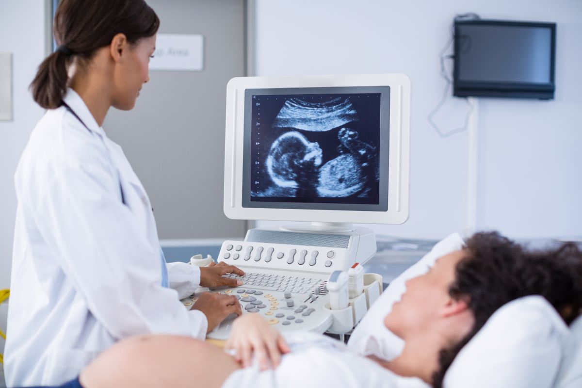
When you’re pregnant, your doctor will recommend you come in regularly for ultrasounds (also called a sonogram). These are tests that use high-frequency sound waves to capture the fetus developing inside of you, and offer a chance for both doctor and parent to examine them.
While it is more commonly known as a way that parents get their first look at their baby, ultrasounds have many more purposes and are a critical part of a developing pregnancy. If you’re wondering what they are, here is the importance of ultrasound in pregnancy, and how it helps in the health of a growing baby.
Confirms the pregnancy


Home pregnancy tests are often how many women find out they may be pregnant — however, these tests aren’t 100% accurate. An ultrasound, as well as a medical pregnancy test administered by a healthcare professional, is the best way to confirm a possible pregnancy.
Diagnoses an ectopic pregnancy or miscarriage
An ectopic pregnancy is what happens when a fertilized egg or fetus doesn’t attach itself to the uterus — instead lodging somewhere else, like the fallopian tube. An ultrasound is critical for diagnosing this problem, as it is not often apparent through its symptoms. Early diagnosis is important as the patient will need immediate medical attention to remove the fetus. If you find you’re dealing with an ectopic pregnancy, your doctor will discuss possible treatments and the best course of action for you.
Estimates due date


Normal pregnancies take around 37 to 41 weeks to complete. You must be able to track how far along you are in your pregnancy so that you can compare your progress to well-established growth charts.
Early in a normal pregnancy, an ultrasound is one of the most accurate ways to measure growth. Thus, it is also a great tool in estimating your due date. An ultrasound helps your doctor measure your baby’s length from crown to rump. This measurement helps them estimate how far along you are, and see if you are developing normally. Knowing your due date will also ensure your baby is not delivered too early, or too late.
Checks for multiple pregnancies
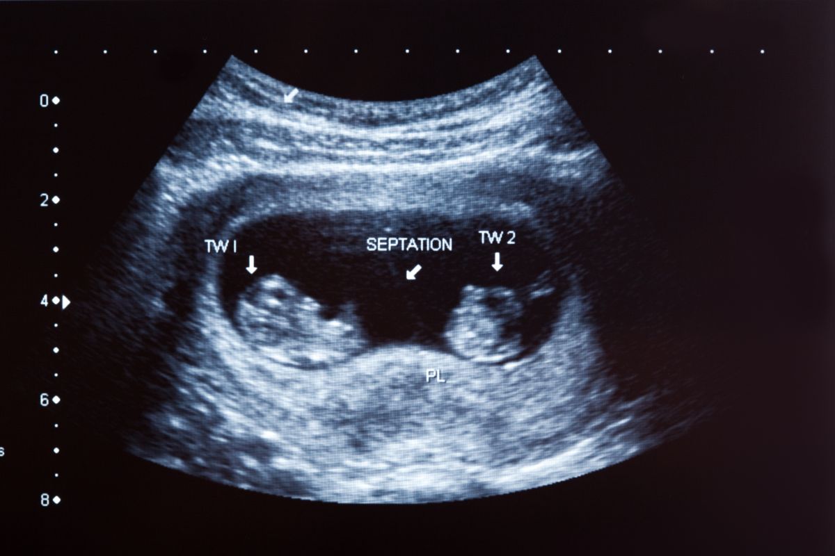
Ultrasounds can detect your baby’s heartbeat — and show if there is more than one set of heartbeats. If your doctor detects multiple heartbeats, you will likely be having twins, triples, or more babies in this pregnancy.
Carrying more than one baby has special risks, which must be monitored by your doctors regularly. This will enable them to promptly treat you if complications like twin-to-twin transfusion occur.
Identifies problems with the placenta
The condition and position of the placenta are crucial to your baby’s health and are one of the things your doctor needs to monitor carefully. An ultrasound can determine if there are any issues with your placenta, such as:
Placenta Previa
This is when the placenta lies unusually low in your uterus, usually next to or covering the cervix. It’s not uncommon for your placenta to be close to the cervix and gradually move upwards the uterine wall as your pregnancy progresses. However, if your placenta is still lying very low late into the pregnancy, or even during labor, it can result in extensive bleeding, injuring both mother and child.
An ultrasound is crucial in determining if you are dealing with Placenta Previa, which shows your doctor that delivery is best done via a cesarean section to avoid complications.
Vasa Previa
This is when the baby’s blood vessels run near or cross over the cervix. These blood vessels are unsupported by their placental tissue or umbilical cord, which increases the risk of rupturing when your membranes rupture during delivery.
Ultrasounds that identify a Vasa Previa may require you to be hospitalized for a prolonged period, and to birth via cesarean section to ensure safe delivery.
Placenta Accreta
This happens when the blood vessels and other parts of the placenta grow too deeply into the uterine wall, which makes detaching the placenta during childbirth difficult. When the placenta remains firmly attached after delivery, it can cause severe bleeding.
Placenta Increta
Similar to Placenta Accreta, this condition happens when parts of the placenta grow and invade the muscles of the uterus.
Placenta Percreta
This condition is when the placenta grows into and past the uterine wall.
Checks for congenital anomalies
Many parents are concerned if their baby is growing healthily or may be experiencing genetic problems. An ultrasound can help your doctor identify and diagnose possible congenital anomalies. The earlier your doctor can do so, the more time you will have to make a decision regarding your pregnancy, or to prepare for the difficulties associated with the particular condition.
Monitors fetal growth
An ultrasound at 20 weeks is generally recommended for all pregnancies, but most doctors will ask that you come in regularly so that they can monitor fetal growth at critical stages.
Doing so gives your doctor a clearer picture of how well your baby is growing. If their growth does not match up with expected norms, this can indicate problems with either the placenta or your baby. Early identification and intervention are key to dealing with many fetal conditions.
Ultrasounds can also show you the gender of your baby after 20 weeks. Note that ultrasounds are not always 100% accurate, as the ultrasound images can be misinterpreted.
Monitors fetal position
The baby’s position (breech, transverse, cephalic, or optimal) in your uterus needs to be monitored during the days leading to your delivery. Their position needs to be known, as it affects which method of delivery is most appropriate for your pregnancy.
Monitors amniotic fluid
Amniotic fluid is a substance produced by a developing fetus. Too much or too little amniotic fluid can be a symptom of pregnancy problems. Ultrasounds can monitor the level of amniotic fluid in your uterus, which helps in ensuring prompt intervention or medical care when problems here occur.
Key Takeaway
The importance of ultrasound in pregnancy lies in its diagnostic functions, allowing doctors and other healthcare professions to identify possible issues early on. For most pregnancies, an ultrasound is a safe and effective way to monitor your baby’s development and ensure a healthy pregnancy and delivery.
If you want only the best care for you and your growing baby, work with the doctors at Perpetual Health Medical Center — Las Pinas (PHMC). Our experienced doctors can meet your pregnancy needs. We also offer Maternity Packages available for Normal and Caesarean Delivery. Learn more about it by sending us a message here.