


Emergency / Hotline: 8874-8515

Institute of Radiology and Allied Sciences

Get fast, accurate imaging results using advanced MRI, CT, and diagnostic technologies.

About Institute of Radiology and Allied Sciences

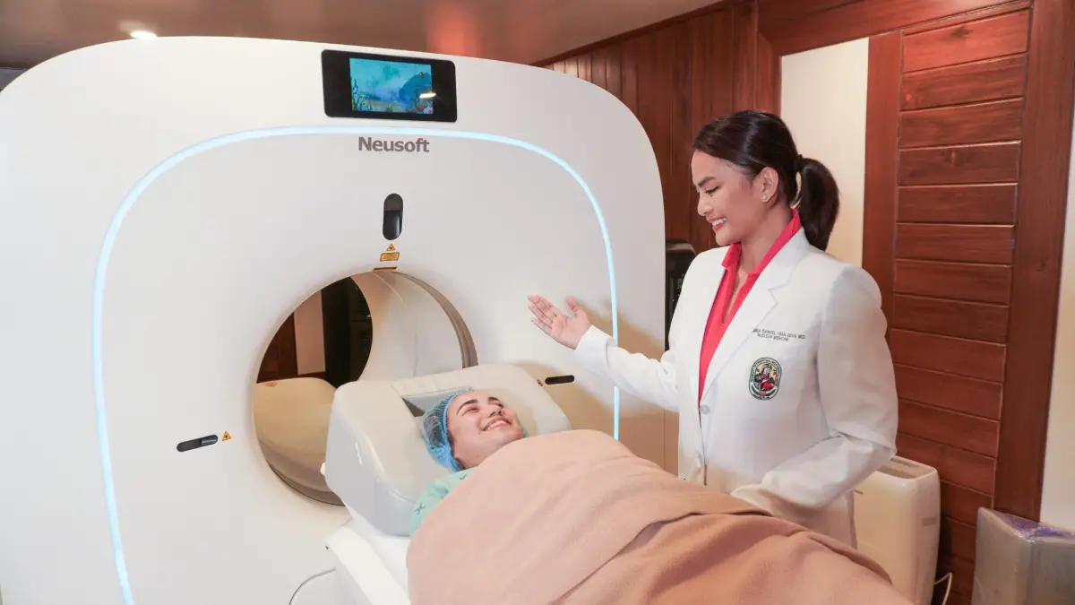
The Institute of Radiology and Allied Sciences is dedicated to delivering comprehensive diagnostic and interventional imaging services with precision, efficiency, and care. Equipped with advanced technologies such as X-ray, ultrasound, CT scan, MRI and interventional radiology suites, the Institute plays a vital role in early detection, accurate diagnosis, and effective treatment planning.

Our team of expert radiologists, technologists, and allied health professionals work closely with physicians across specialties to provide timely, reliable results. Committed to patient safety, comfort and innovation, we strive to set the standard in imaging excellence.
Gallery

Share our Institute of Radiology and Allied Sciences

Facebook
LinkedIn
Email
X
Main Line

(632) 887488515

📱
Mobile

Viber: 095177744081

📍
Location

GF Main Hospital Building, Wing A

OUR CENTERS & FACILITIES

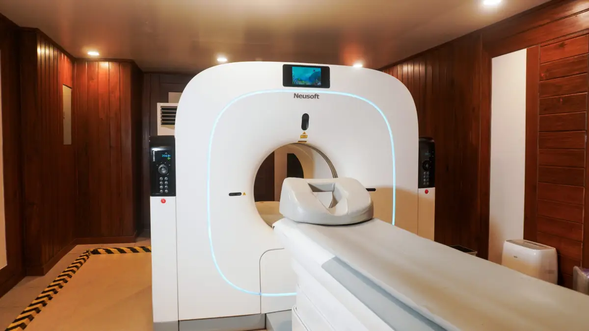
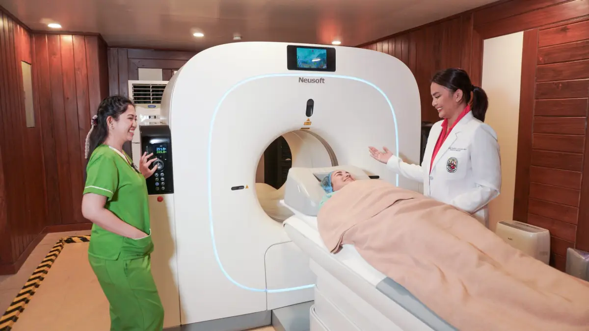
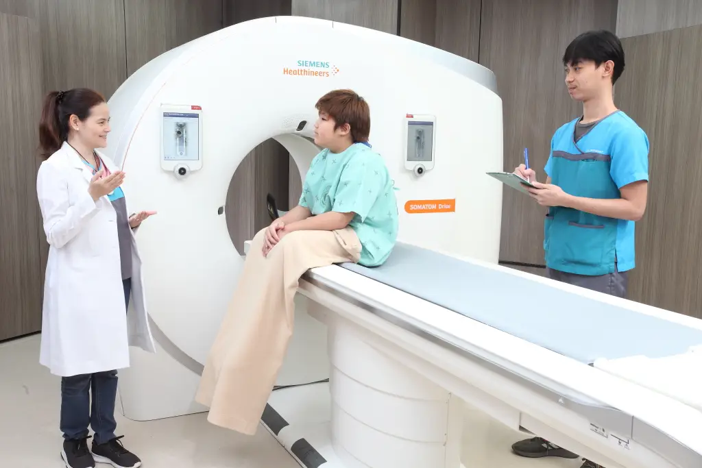
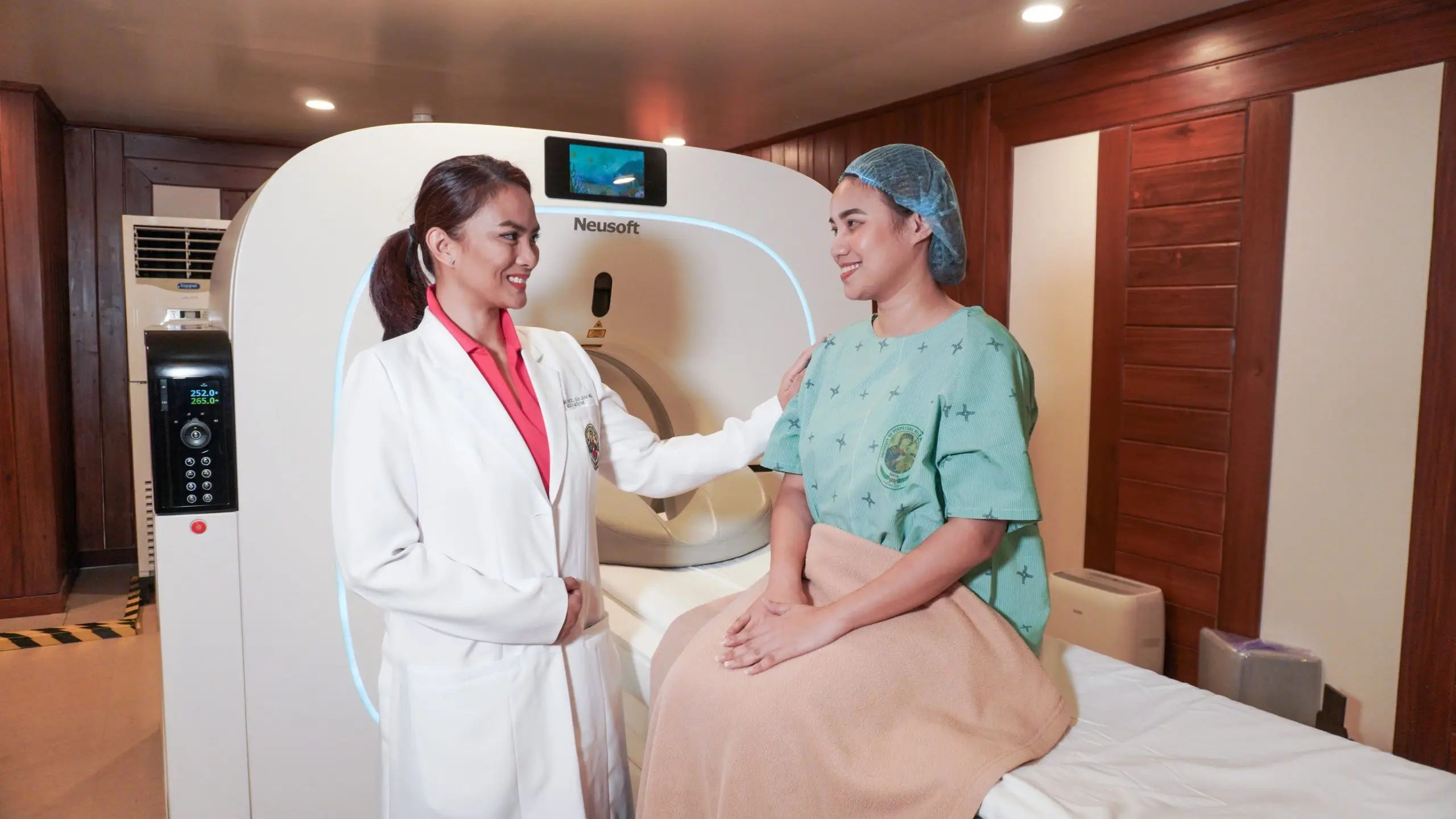
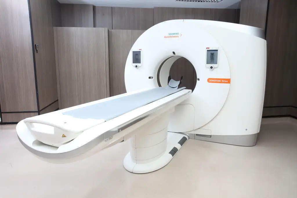
Our hospital is equipped with 2 CT scans, one of which is the 768-slice SOMATOME Drive Dual Source CT-Scan. This CT-Scan makes use of dual-source technology (2 X-ray tubes and detectors) to provide fast, high-resolution imaging with reduced radiation exposure.

View Service

Our Bone Densitometry Unit specializes in measuring bone mineral density to assess bone strength and detect conditions like osteoporosis. Using advanced dual-energy X-ray absorptiometry (DEXA) technology, the test is quick, painless, and uses very low radiation. Results help doctors evaluate fracture risk, monitor bone health, and guide treatment plans. We provide accurate, reliable testing in a safe and comfortable environment.

View Service

Elastography is a specialized ultrasound technique that measures the stiffness or elasticity of tissues, helping detect and evaluate conditions such as liver fibrosis, tumors, or other abnormalities. It is non-invasive, painless, and provides valuable information that can guide diagnosis and treatment without the need for a biopsy in many cases. Our elastography services use advanced technology to deliver accurate results quickly and comfortably.

View Service

Fluoroscopy is a specialized imaging technique that uses a continuous X-ray beam to create real-time moving images of the body. It is often used to guide diagnostic tests and treatments, such as barium studies, catheter placements, or joint injections. This dynamic imaging method allows doctors to see organ movement, track contrast agents, and perform procedures with precision. Our fluoroscopy services use modern, low-dose equipment to ensure patient safety while providing clear, accurate results.

View Service

General X-ray, or radiography, is a quick and painless imaging technique used to examine bones, chest, and other parts of the body. It works by passing a small dose of radiation through the body to create detailed images that help detect fractures, infections, or abnormalities. Our radiology team uses modern, low-dose equipment to ensure patient safety while delivering accurate results for prompt diagnosis and treatment.

View Service

General X-ray, or radiography, is a quick and painless imaging technique used to examine bones, chest, and other parts of the body. It works by passing a small dose of radiation through the body to create detailed images that help detect fractures, infections, or abnormalities. Our radiology team uses modern, low-dose equipment to ensure patient safety while delivering accurate results for prompt diagnosis and treatment.

View Service

General or whole body ultrasound is a safe, painless imaging procedure that uses high-frequency sound waves to produce real-time images of organs, tissues, and blood flow. It is commonly used to assess the abdomen, pelvis, thyroid, blood vessels, and other soft tissues, and can also aid in guiding certain medical procedures. Because it does not use radiation, ultrasound is suitable for patients of all ages. Our skilled sonographers and radiologists ensure accurate results in a comfortable, patient-centered environment.

View Service

Interventional radiology uses advanced imaging guidance—such as X-ray, ultrasound, or CT—to perform minimally invasive procedures for diagnosis and treatment. These techniques often replace the need for traditional surgery, resulting in smaller incisions, less pain, and faster recovery times. Common procedures include angioplasty, stent placement, image-guided biopsies, and targeted cancer treatments. Our interventional radiology team works closely with other specialists to provide precise, safe, and effective care tailored to each patient’s needs.

View Service

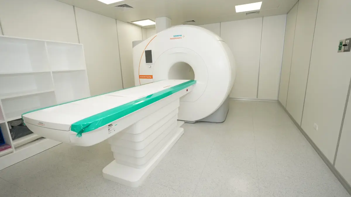
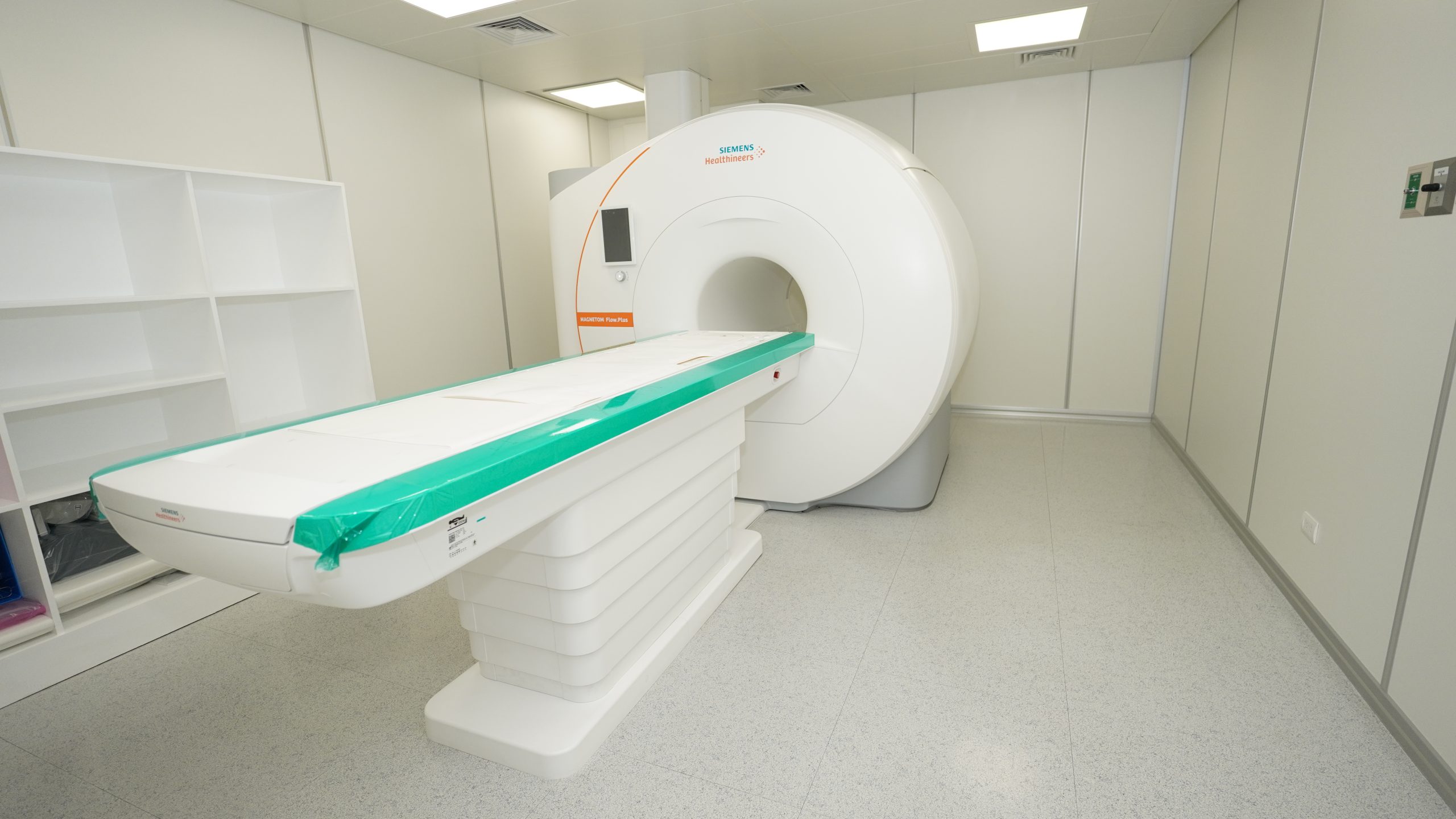
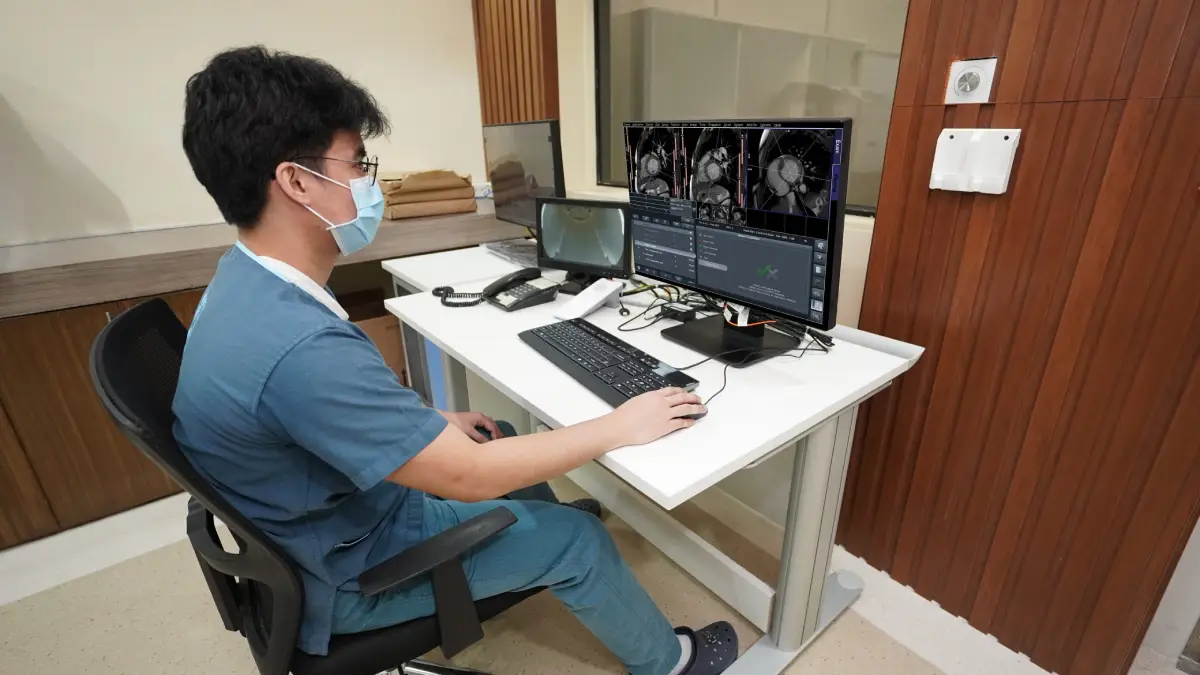
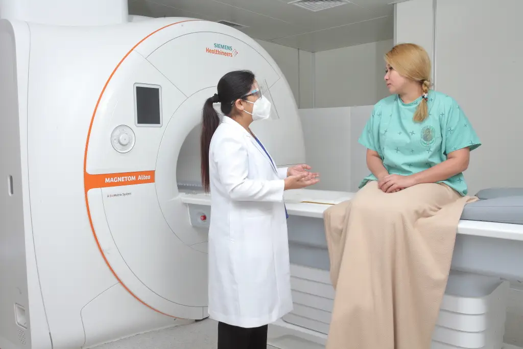
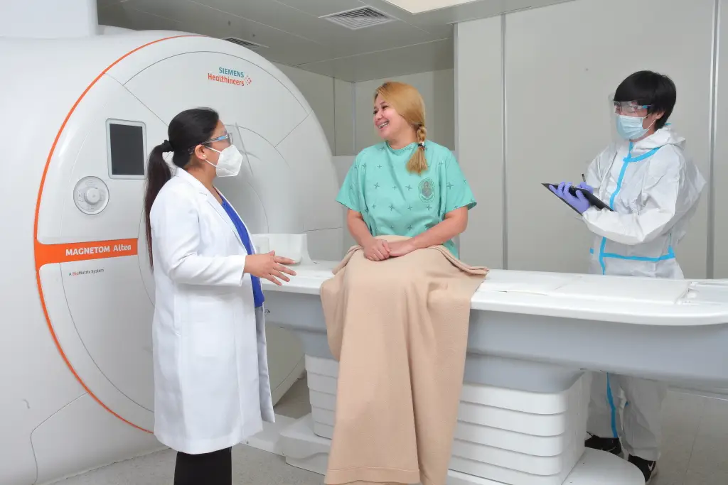
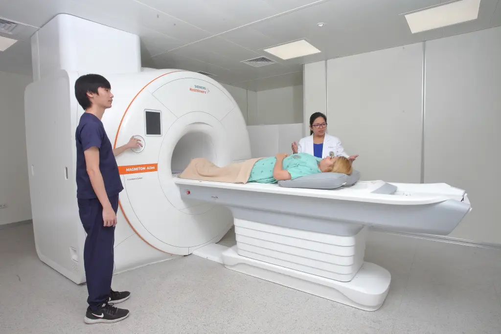
Patients often hesitate to get MRI scans because of long wait times, discomfort, and anxiety inside the machine. PHMC addresses that head-on by investing in newer, smarter MRI systems that focus on speed, comfort, and accuracy. MAGNETOM ALTEA The MAGNETOM Altea is built around one simple idea: make MRI less stressful for patients while giving doctors better images to work with. At the core of this system is BioMatrix technology, which adapts the scan to each patient’s body automatically. Instead of relying on manual adjustments, the machine uses sensors and smart positioning coils to detect anatomy and breathing patterns. That means fewer setup errors, more consistent image quality, and less need to repeat scans. It also uses Tim 4G (Total Imaging Matrix), which allows multiple coils to work together seamlessly. This speeds up the entire scanning process and enables full-body imaging without constantly repositioning the patient. For patients, this translates to shorter scan times and less time inside the machine. Comfort is a big upgrade here. The system is designed to reduce motion sensitivity, so even patients who have difficulty staying still—like elderly individuals or those in pain—can still get clear results. The quieter operation and more open feel also help reduce anxiety. For doctors, the benefit is straightforward: clearer, more consistent images delivered faster. That leads to better diagnosis and quicker clinical decisions. MAGNETOM FLOW The MAGNETOM Flow focuses on sustainability and reliability without compromising diagnostic quality. This system uses DryCool technology, making it virtually helium-free. Traditional MRI machines rely heavily on liquid helium for cooling, which is expensive, limited, and requires complex infrastructure. By minimizing helium use, Magnetom Flow reduces operational costs and environmental impact. For the hospital, this means fewer disruptions and lower maintenance requirements. For patients, it means more consistent availability of MRI services—less downtime, fewer cancellations, and faster scheduling. Despite being more efficient, it still delivers high-quality imaging across a wide range of clinical applications, from neurological scans to musculoskeletal and abdominal imaging. It’s designed to handle everyday diagnostic demands while maintaining stable performance. What This Means for Patients at PHMC At Perpetual Help Medical Center, having both systems in one facility creates a strong advantage. Patients benefit from: Doctors benefit from: In simple terms, PHMC’s MRI capabilities are built to remove friction—from patient anxiety to technical limitations—so imaging becomes faster, smoother, and more dependable.

View Service

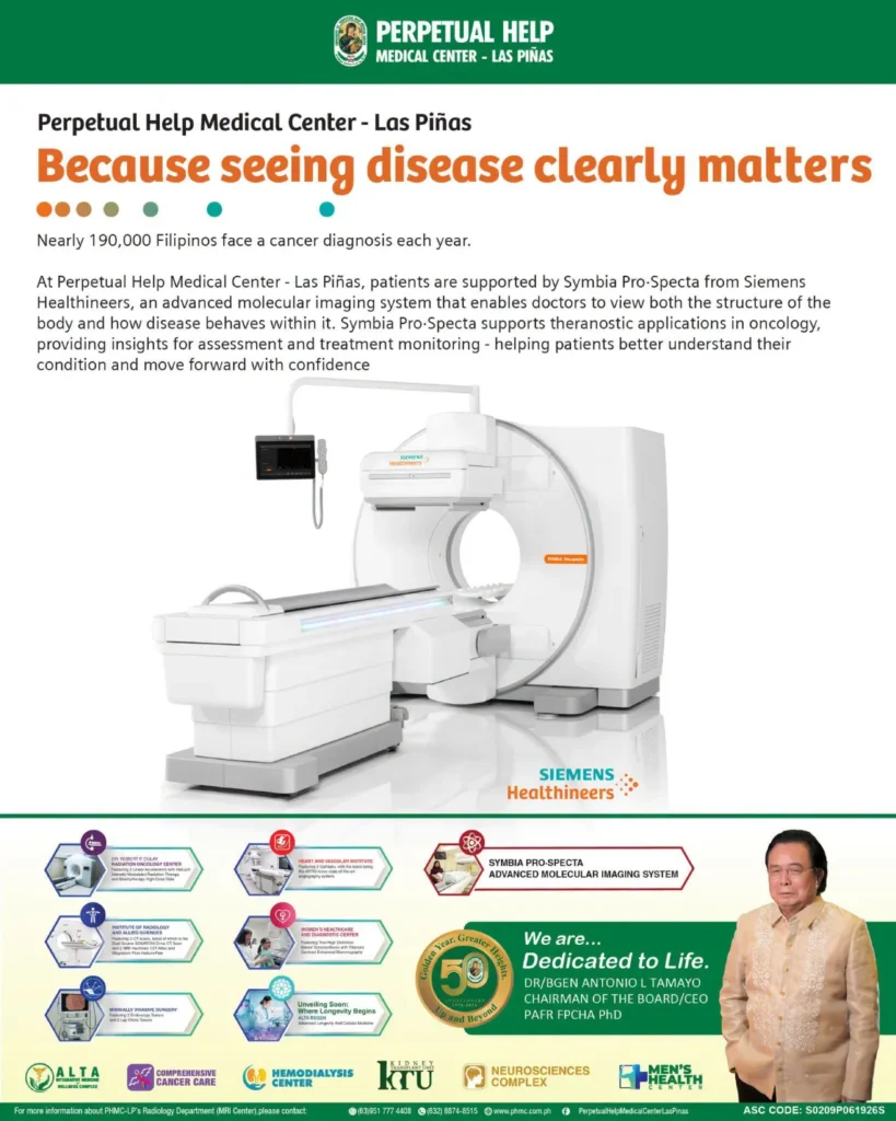
PET/CT scan combines two advanced imaging technologies—PET (Positron Emission Tomography), which shows how organs and tissues are functioning, and CT (Computed Tomography), which provides detailed images of their structure. This powerful combination allows doctors to detect cancer, evaluate heart and brain disorders, and monitor treatment response with high accuracy. Our PET/CT services use the latest

View Service

PET/CT scan combines two advanced imaging technologies—PET (Positron Emission Tomography), which shows how organs and tissues are functioning, and CT (Computed Tomography), which provides detailed images of their structure. This powerful combination allows doctors to detect cancer, evaluate heart and brain disorders, and monitor treatment response with high accuracy. Our PET/CT services use the latest

View Service

Ultrasound scanning, or sonography, uses high-frequency sound waves to create real-time images of the body’s internal structures. It is a safe and painless procedure that does not involve radiation, making it ideal for patients of all ages, including expectant mothers. Ultrasound is widely used to examine the abdomen, pelvis, heart, blood vessels, and other soft tissues, as well as to guide certain medical procedures. Our team ensures accurate, timely results while prioritizing patient comfort.

View Service