The Perpetual Help Medical Center (PHMC) respects the privacy rights of all
individuals and is committed to handling personal data responsibly and in accordance with
the Republic Act 10173, known as the Data Privacy Act of 2012, and its pertinent rules and
regulations, because your privacy is important to us.
This is not a consent form but this is PHMC’s general statement on its data
processing activities to notify Data Subjects of Personal Data processed and the purpose and
extent of processing. PHMC may further provide other notices at the time of
specific activities.
PHMC collects the following data: 1) Personal details- name, birth, gender,
civil status and affiliations; 2) Contact information- address, email, mobile and telephone
numbers; 3) Medical information- physical, psychiatric and psychological information;
4)Employment information- government-issued numbers, position and functions; 5)Applicant
information- academic background and previous employments; 6) Academic information- grades,
course and academic standing; 7) Supplier and 3rd Party Provider information- company
profile, DTI/ SEC registration, business permits and licenses, BIR registration and other
business related information. PHMC may likewise collect other information that it believes
are relevant to meet the requirements of government authorities and for any other legitimate
purposes.
PHMC processes data to: 1) Conduct its functions, perform its obligations,
and exercise its rights as a healthcare service provider; 2) Act for the holistic welfare of
patients, service recipients, and their respective representatives and companions; and, 3)
Manage its affairs as a company, medical, and educational institution with its own
obligations and rights.
The PHMC Website uses cookies to personalize the user’s browsing experience, link to
social media sharing, troubleshoot issues, and monitor site visits and NOT in any way
collect personal information for any form of processing.
PHMC collects Personal Data through submission by the Data Subject and by
affiliates through electronic systems and platforms, e-forms, email, or through printed
forms, attachments, and other documents required by its medical and administrative offices
at the onset of service, transaction or processing. With regard to personal data from
affiliates, PHMC maintains a Data Sharing Agreement particularizing the obligations of the
parties thereto foremost of which is its protection and privacy.
PHMC collects Personal Data from Patient at entry points or upon
registration at Inpatient, Outpatient and ER departments.
PHMC stores and protects data in physical and electronic forms: Managed by
its medical and administrative offices, physical records in folders/ envelopes are
ultimately stored in box files in shelves in a dedicated secured area while electronic
records are stored in secured servers with high availability and redundancy.
PHMC collects and uses Personal Data proportionately as necessary for its
legitimate purposes in providing best service to patients in accordance with the PHMC
policies and in compliance with the DPA of 2012 and requirement of the Department of Health
and other government bodies.
PHMC retains data in accordance with its policies on retention compliant to
government rules and regulations, such as but not limited to, those from the Bureau of
Internal Revenue (BIR), the Department of Health (DOH), the Philippine Health Insurance
Corporation (Philhealth), the Local Government Units (LGUs), among others.
Contact:Contact: Inquiries and concerns on data privacy may only be
directed to PHMC Data Protection Office: PHMC
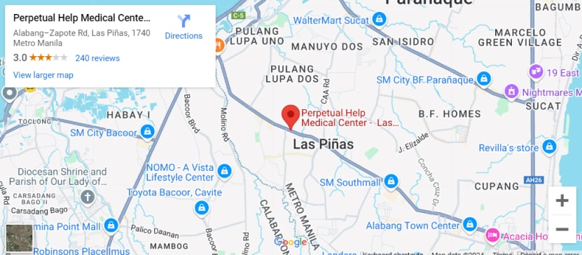
Address: Alabang-Zapote Road, Pamplona 3, Las Pinas 1704
Telephone: 8874-8515